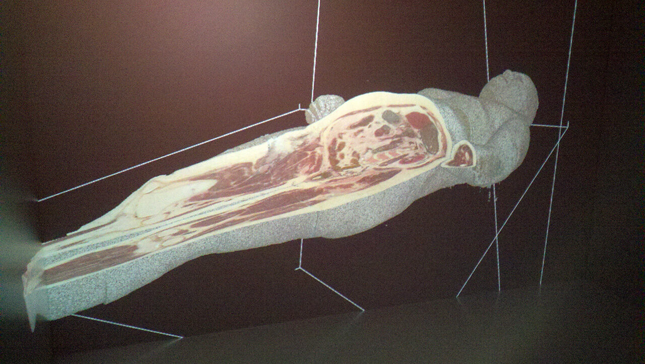
The 3-D cadaver floats in space like a hologram on an invisible gurney.
University of Michigan 3-D Lab employee Sean Petty stands a few inches away. He wears special glasses and uses a joystick to arbitrarily slice away layers of the virtual-reality cadaver, revealing deep red muscle tissue. He enlarges and turns the body for a better view of the detailed anatomy inside.
Alexandre DaSilva, assistant professor at the School of Dentistry, says working with the virtual-reality cadaver is the opportunity of a lifetime for himself and for his dental students, residents, and doctoral students.
“The first time I saw the technology I almost cried,” says DaSilva, who also heads the Headache and Orofacial Pain Effort at U-M Dentistry and the Molecular and Behavioral Neuroscience Institute. “In my wildest dream, I never thought this would be possible.”
The 3-D model supplements traditional anatomy class in several ways, DaSilva says. For instance, the residents can back up and redo cuts, and also enlarge areas to see them more closely.
“When you really immerse in the 3-D image, you can use all your senses,” says Thiago Nascimento, a postdoctoral student in DaSilva’s research lab. “It blew my mind.”
Researchers at the U-M Medical School Visible Human Project had already layered the cadaver into very thin sections. The group shared the anatomical data with Petty and 3-D Lab colleague Theodore Hall. The two then wrote the code that reassembled the body into a computer model, says Eric Maslowski, manager of the 3-D Lab, which is part of the Digital Media Commons, a service of the library.
Maslowski says other disciplines, such as engineering or natural science, can use the technology to virtually dissect simulated hurricanes or slice into Mastodons, for example. DaSilva uses it now to study 3-D brains of pain patients to learn about migraine and other disorders.
Top image: Dental residents virtually dissect this 3-D cadaver, which is anatomically correct. (Photo: Laura Bailey.)


Bob Kennedy - 2002
So does this have any diagnostic application for live subjects, or patients? Or should I ask how soon might this be the case?
Sounds like this could give the ability to explore a person’s anatomy before surgery or other treatments for a better diagnosis.
Nice.
Reply