Going viral
You’ve seen the image — a large spherical mass with protruding red spikes plastered across the television and the internet. It’s an image that has become all too familiar over the last few months.
We know it well as COVID-19, but what exactly does the image tell us other than serving as the visual representation of the pandemic? Deb Gumucio, co-founder and director of U-M’s BioArtography Project has thought a lot about the impact scientific imagery has when understanding biological diseases, viruses, and the human body in the time of the coronavirus.
“Right now, the image that everyone sees is essentially being treated like a brand,” says Gumucio, also a professor emerita of cell and developmental biology at the U-M Medical School. “A lot of people look at this image and they don’t have a lot of information about it — it looks a little like some sort of menacing alien machine.”
The beauty of biology

“Family Tree” by Greggory Myers, Graduate Student, Cell and Developmental Biology (Khoriaty Laboratory), Department of Internal Medicine, University of Michigan Medical School
Gumucio co-founded BioArtography as a fundraising initiative for U-M research labs in 1995, but over the last 25 years, it has emerged as something far greater: a way to educate the public about groundbreaking scientific research and technologies occurring at Michigan Medicine and beyond.
Gumucio and her team take microscopic images from current and past biological research, and a committee composed of U-M scientific researchers and professors at the U-M Stamps School of Art & Design select the most striking, dynamic images to display and sell. Proceeds from the sale of the work help support the training of our next generation of researchers.
“You’ve probably never seen your pancreas, or brain, or cells under the microscope, and if you did without knowing what it was, they would probably look a bit like an abstract blob,” Gumucio says. “That’s because when you first look under a microscope at cells and tissues, you don’t see much of anything because cells and tissues are transparent for the most part, with the exception of plant cells and tissues.”

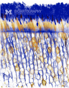
“Seeing the Light” by Jillian Pearring, Ph.D., Assistant Professor, Department of Ophthalmology and Visual Sciences, University of Michigan Medical School
According to Gumucio, to see tissues and cells under the microscope, researchers have to stain them with color. There are many common stains; for example, a blue color stain is often used to indicate a cell nucleus, and a red stain is commonly used for fats. These stains, and sometimes, added color and modifications in Photoshop, make the colorful images offered by Bioartography.
BioArtography’s images are symmetrical, colorful, and strikingly shaped. Yet beyond the color and artistic form, the shapes and figures that make up the image are extremely important in understanding how the human body functions. That’s precisely the goal of BioArtography—to demonstrate that these powerful, beautiful images also can be used as a teaching tool.
Gumucio says that it is easier to talk to nonscientists about complex biology and research using visuals.
“People may be fearful when they first hear about technologies, such as human embryonic stem cells, or organoids, but the images produced at BioArtography makes this science approachable,” she says. “The art it creates is an entryway into science. For example, when you look at the image of the coronavirus, you might think, ‘What are those spikes on the surface? How does this tiny thing make us sick?’”
Coronavirus: Decoding the spiky fuzzball
How does that famous image of the coronavirus, created by the medical artists at the Center for Disease Control and Prevention, compare to the work done by BioArtography?

“Nasty Noro” BioArtography image. Noroviruses have a similar overall structure to coronaviruses, but noroviruses are molecularly very distinct. Noroviruses cause the majority of viral-induced diarrhea/vomiting in places where crowds gather.
The popular coronavirus image is actually a digital reproduction. While the images that Gumucio creates as part of her BioArtography process are from actual photo-micrographs, she says that the image we’ve seen circulating is a helpful visual tool.
“It gives you a fairly accurate picture of the way the virus is put together,” she says. “Those spikes on the outside are really there and they’re what gave the virus its name—’corona’ comes from the word crown in Latin, and those spikes bind to the receptor on a cell and allow the virus to enter it.”
Like each image that BioArtography produces, the coronavirus’ spiky ball tells a story, and that story has become an important visual cue about public health and safety.
“In the image, they added some orange and yellow-colored dots on the surface, which represent the myriad of proteins that the virus encodes,” Gumucio says. “That’s why washing your hands with soap is so important — it denatures those proteins on the surface. If the virus does not have those proteins, it cannot infect a cell.”
She notes that the digital microscopic rendering of the virus is a good thing, and could actually serve as a constant public health reminder.
“Though the image doesn’t often come with an explanation, people should know that it is a visual representation of how they have the power to destroy those proteins if they wash their hands thoroughly,” she says.
According to Gumucio, the problem with the coronavirus image is that it is being treated as a brand for the virus, and not as the educational tool that it should be.
“I’d like to see more captions and more people talking about what the image can actually tell us,” she says. “BioArtography promotes the idea that every single image that we generate is unique and tells a unique story about the image researched.”
Gumucio wants us to think about the image in a different way the next time we see it.
“There is a lot of fear about the virus and the image that represents it,” she says. “If people had more information to help them understand it, I think they’d feel a bit more confident that there are ways to defeat it.”
(Top Photo Credit: CENTERS FOR DISEASE CONTROL AND PREVENTION HANDOUT/EPA-EFE/REX (10544648b); an undated handout photo made available by the Centers for Disease Control and Prevention (CDC) in Atlanta, Ga., (issued 31 January 2020) of an illustration created at the CDC revealing ultrastructural morphology exhibited by the 2019 Novel Coronavirus (2019-nCoV). Note the spikes that adorn the outer surface of the virus, which impart the look of a corona surrounding the virion, when viewed electron microscopically. This virus was identified as the cause of an outbreak of respiratory illness first detected in Wuhan, Hubei Province, China. U.S. national public health institute releases illustrations revealing structure of novel coronavirus, Atlanta, USA – 31 Jan 2020).


Jyotirmoy SARKAR - 1990, PhD in Statistics
I studied the spikes from a mathematician’s point of view; how many are there? where are they located? I believe a sculpture of the spikes on the surface of the sphere can be made, serving as an educational tool. Should you be interested, I will be happy to send you a copy of the paper. Thank you.
Reply
Karen Boucek Senn - 1971
AWESOME! I applaud your work! Thank you for sharing! (I graduated from L S & A in physical therapy.)
Reply
Julie Bender - BFA 1981
As an artist, I really appreciate your work, because the visuals communicate so strongly with me. I will remember the orange and yellow dots representing the myraid of proteins as I wash my hands. Thank you!
Reply